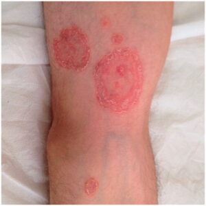
Pitryiasis rosea is a somewhat common distinctive looking rash with a self-limited course that predominantly affects children and young adults. It typically starts with a large oval to round salmon-red patch with a fine, wrinkled tissue like scale trailing behind the border.
Within one to two weeks of this “herald patch”, smaller, similar oval shaped lesions erupt over the trunk and proximal extremities. Numerous lesions on the back sometimes give the characteristic drooping pine tree or “Christmas tree distribution”.
PR is now felt in most cases to be a reactivation of a childhood virus. The rash is typically asymptomatic and clears in about 6-8 weeks on its own. Occasionally it can last longer, and itching can become more prominent with more extensive involvement. It also can be misdiagnosed as other similar appearing skin conditions and mistreated; so consultation with a Dermatologist can be reassuring

Background: Pityriasis rosea (PR) is a common, acute, self-limiting inflammatory skin disease. It can easily be recognized with its typical clinical presentation. However, unusual clinic presentations can cause difficulty in diagnosis. Up to now, not many atypical forms are reported.
Objective: To determine the clinical characteristics of patients with atypical pityriasis rosea.
Methods: A total of 27 cases, diagnosed as atypical PR by clinical and/or histopathological examination and applied to the outpatient clinic of dermatology department between the years 2007 and 2015 were analyzed retrospectively.
Results: The ages of patients ranged from 2 to 59 years. Of these patients, 15 (55.6%) were male and 12 (44.4%) were female. The male-to-female ratio was 5–4. Five patients had papular, four patients had purpuric, three patients had vesicular, two patients had follicular, one patient had erythema multiforme-like and one patient had eczematous drug-induced atypical form of pityriasis rosea. There were 12 cases of localized, two cases of segmental pityriasis rosea. Four of the localized forms also had atypical morphology. Histopathological evaluation was required for diagnosis in 12 (44.4%) patients.
Conclusions: PR can appear in many different uncommon forms. Localization and skin rush can be misleading and diagnosis can be compelling.